Effect of Spatial, Intensity, and Hybrid Augmentation on Kidney CT Image Classification
DOI:
https://doi.org/10.56705/ijodas.v7i2.443Keywords:
Kidney CT Images, Data Augmentation, Intensity Augmentation, Deep Learning, Medical Image Classification, Learning Behavior AnalysisAbstract
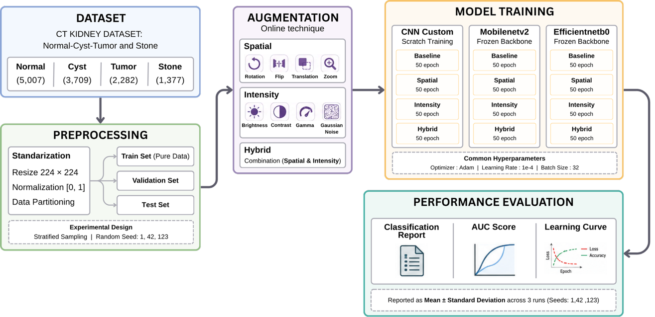
Introduction: Kidney diseases remain a major global health challenge, and computed tomography (CT) provides detailed renal imaging that supports accurate diagnosis. Although data augmentation is commonly used to improve deep learning performance on limited medical datasets, the effects of different augmentation strategies on image characteristics and model learning behavior remain insufficiently understood. Method: This study evaluated spatial, intensity, and hybrid augmentation for kidney CT image classification using a custom CNN, MobileNetV2, and EfficientNet-B0. A public dataset containing 12,446 CT images across Normal, Cyst, Tumor, and Stone classes was partitioned using stratified sampling into training, validation, and test sets. Four scenarios—baseline, spatial, intensity, and hybrid augmentation—were evaluated across three independent random seeds using accuracy, precision, recall, F1-score, and AUC. Results and Discussion: The baseline achieved mean accuracies of 99.96%, 98.32%, and 94.06% for CNN, MobileNetV2, and EfficientNet-B0, respectively. Intensity augmentation slightly improved CNN accuracy to 99.99% and consistently produced smaller performance degradation and more stable convergence than spatial and hybrid augmentation. Spatial and hybrid transformations generally reduced classification performance, indicating that excessive geometric changes may disrupt diagnostically relevant anatomical features. Conclusion: Baseline training provided the best overall performance, while intensity augmentation was the most effective augmentation strategy, demonstrating that preserving anatomically meaningful image characteristics is more important than indiscriminately increasing data diversity.
Downloads
References
[1] A. Francis et al., “Chronic kidney disease and the global public health agenda: an international consensus,” Nat. Rev. Nephrol., vol. 20, no. 7, pp. 473–485, Jul. 2024, doi: https://doi.org/10.1038/s41581-024-00820-6.
[2] R. Setyati et al., “The Importance of Early Detection in Disease Management,” Journal of World Future Medicine, vol. 2, no. 1, pp. 51–63, 2024, doi: https://doi.org/10.55849/health.v2i1.692.
[3] S. S. R. Vulasala et al., “Computed tomography of renal emergencies: A comprehensive diagnostic guide for radiology residents,” World J. Nephrol., vol. 15, no. 1, Mar. 2026, doi: https://doi.org/10.5527/wjn.v15.i1.114185.
[4] I. D. Mienye, T. G. Swart, G. Obaido, M. Jordan, and P. Ilono, “Deep Convolutional Neural Networks in Medical Image Analysis: A Review,” Information, vol. 16, no. 3, p. 195, Mar. 2025, doi: https://doi.org/10.3390/info16030195.
[5] H. E. Kim, A. Cosa-Linan, N. Santhanam, M. Jannesari, M. E. Maros, and T. Ganslandt, “Transfer learning for medical image classification: a literature review,” BMC Med. Imaging, vol. 22, no. 1, p. 69, Dec. 2022, doi: https://doi.org/10.1186/s12880-022-00793-7.
[6] H. Khachnaoui, B. Chikhaoui, N. Khlifa, and R. Mabrouk, “Enhanced Parkinson’s Disease Diagnosis Through Convolutional Neural Network Models Applied to SPECT DaTSCAN Images,” IEEE Access, vol. 11, pp. 91157–91172, 2023, doi: https://doi.org/10.1109/ACCESS.2023.3308075.
[7] G. Tummalapalli, O. Gurrapu, K. N. Kumar, J. Venkata Suman, A. V. Rao, and M. Prabhu, “Deep Learning Approaches for Enhancing Image Classification Accuracy in Medical Imaging,” in 2025 Devices for Integrated Circuit (DevIC), IEEE, Apr. 2025, pp. 16–21. doi: https://doi.org/10.1109/DevIC63749.2025.11012289.
[8] J. C. L. Chow, “Machine learning in cancer imaging for enhanced precision in diagnosis and therapy,” Discover Computing, vol. 29, no. 1, p. 186, Mar. 2026, doi: https://doi.org/10.1007/s10791-026-10078-0.
[9] F. Jahan, A. S. Reza, M. K. Morol, D. Nandi, Md. J. Hossen, and M. Rahman, “Explainable deep learning for early diagnosis of chronic kidney disease from CT images in Bangladeshi patients,” Sci. Rep., vol. 16, no. 1, p. 14819, Mar. 2026, doi: https://doi.org/10.1038/s41598-026-42654-1.
[10] Y. Dang et al., “Data Augmentation for Sequential Recommendation: A Survey,” IEEE Trans. Knowl. Data Eng., vol. 38, no. 8, pp. 4938–4957, Aug. 2026, doi: https://doi.org/10.1109/TKDE.2026.3692969.
[11] N. Kozah, F. Dornaika, J. Charafeddine, and J. El Jaam, “Data Augmentation Techniques for Medical Image Segmentation – A Review,” in 2024 International Conference on Computer and Applications (ICCA), IEEE, Dec. 2024, pp. 1–8. doi: https://doi.org/10.1109/ICCA62237.2024.10927851.
[12] F. Garcea, A. Serra, F. Lamberti, and L. Morra, “Data augmentation for medical imaging: A systematic literature review,” Comput. Biol. Med., vol. 152, p. 106391, Jan. 2023, doi: https://doi.org/10.1016/j.compbiomed.2022.106391.
[13] K. Rais, M. Amroune, M. Y. Haouam, A. Benmachiche, and S. Abid, “Dynamic feature context activation and data augmentation for enhanced medical image segmentation,” Multimed. Tools Appl., vol. 85, no. 2, p. 131, Feb. 2026, doi: https://doi.org/10.1007/s11042-026-21296-5.
[14] J. Majidpour and H. Beitollahi, “A Comprehensive Examination of Machine Learning and Deep Learning Approaches for Breast Cancer Detection, Classification, Segmentation, Augmentation, and Feature Selection,” Archives of Computational Methods in Engineering, vol. 33, no. 2, pp. 1913–1944, Mar. 2026, doi: https://doi.org/10.1007/s11831-025-10359-9.
[15] P. Dabove, M. Daud, and L. Olivotto, “Revolutionizing urban mapping: deep learning and data fusion strategies for accurate building footprint segmentation,” Sci. Rep., vol. 14, no. 1, p. 13510, Jun. 2024, doi: https://doi.org/10.1038/s41598-024-64231-0.
[16] D. T. Pham, T. V. Tran, X. Zhu, and H. N. Pham, “Optimising deep learning for building extraction: Dataset efficiency and model backbones under data constraints,” Remote Sens. Appl., vol. 41, p. 101876, Jan. 2026, doi: https://doi.org/10.1016/j.rsase.2026.101876.
[17] M. T. R et al., “Transformative Breast Cancer Diagnosis using CNNs with Optimized ReduceLROnPlateau and Early Stopping Enhancements,” International Journal of Computational Intelligence Systems, vol. 17, no. 1, p. 14, Jan. 2024, doi: https://doi.org/10.1007/s44196-023-00397-1.
[18] S. M. Rayavarapu, T. S. Prasanthi, S. R. Gottapu, and A. Singam, “A Comprehensive Overview on Data Augmentation Techniques for Medical Images,” in 2024 5th International Conference on Electronics and Sustainable Communication Systems (ICESC), IEEE, Aug. 2024, pp. 1324–1329. doi: https://doi.org/10.1109/ICESC60852.2024.10690109.
[19] T. Islam, Md. S. Hafiz, J. R. Jim, Md. M. Kabir, and M. F. Mridha, “A systematic review of deep learning data augmentation in medical imaging: Recent advances and future research directions,” Healthcare Analytics, vol. 5, p. 100340, Jun. 2024, doi: https://doi.org/10.1016/j.health.2024.100340.
[20] S. T. Marc, R. Belavkin, D. Windridge, and X. Gao, “An Evolutionary Approach to Automated Class-Specific Data Augmentation for Image Classification,” 2024, pp. 170–185. doi: https://doi.org/10.1007/978-3-031-50320-7_12.
[21] R. Jain, R. Sharma, D. Tiwari, K. Joshi, and V. Jain, “Enhanced Classification of Intel Images Using Refined EfficientNet and MobileNetV2 Frameworks,” in 2023 4th International Conference on Intelligent Technologies (CONIT), IEEE, Jun. 2024, pp. 1–4. doi: https://doi.org/10.1109/CONIT61985.2024.10627673.
[22] A. Tilevik, “Classification and Performance Metrics,” in Multivariate Statistics and Machine Learning in R For Beginners, Cham: Springer Nature Switzerland, 2025, pp. 147–170. doi: https://doi.org/10.1007/978-3-032-01851-9_10.
[23] Y. Xia and J. Sun, “Area under the Receiver Operating Characteristic Curve (AUC-ROC),” in Machine Learning for Microbiome Statistics, Boca Raton: Chapman and Hall/CRC, 2026, pp. 504–519. doi: https://doi.org/10.1201/9781003610281-17.
[24] O. T. Turan, M. Loog, and D. M. J. Tax, “Generalization performance distributions along learning curves,” Pattern Recognit. Lett., vol. 201, pp. 29–36, Mar. 2026, doi: https://doi.org/10.1016/j.patrec.2026.01.003.
[25] J. Zhu, T. Ma, J. Li, C. Zeng, Q. Fu, and D. Jing, “ETD-Det: Oriented Object Detection With Spectral Diffusion Encoding and Extended-Gaussian Decoding,” IEEE Transactions on Geoscience and Remote Sensing, vol. 64, pp. 1–16, 2026, doi: https://doi.org/10.1109/TGRS.2026.3663195.
[26] S. Krishnendu and M. Biradar, “Enhancing diagnostic information in abdominal computed tomography (CT) images through optimized image enhancement techniques,” Phys. Eng. Sci. Med., vol. 49, no. 1, pp. 475–487, Mar. 2026, doi: https://doi.org/10.1007/s13246-025-01679-y.
Downloads
Published
Issue
Section
License
Copyright (c) 2026 Ardha Ardhana Putra Agustavada, Aji Prasetya Wibawa, Dafa Fadhilah Hilmi, Abdullah Sholum, Felix Andika Dwiyanto

This work is licensed under a Creative Commons Attribution-NonCommercial 4.0 International License.
Authors retain copyright and full publishing rights to their articles. Upon acceptance, authors grant Indonesian Journal of Data and Science a non-exclusive license to publish the work and to identify itself as the original publisher.
Self-archiving. Authors may deposit the submitted version, accepted manuscript, and version of record in institutional or subject repositories, with citation to the published article and a link to the version of record on the journal website.
Commercial permissions. Uses intended for commercial advantage or monetary compensation are not permitted under CC BY-NC 4.0. For permissions, contact the editorial office at ijodas.journal@gmail.com.
Legacy notice. Some earlier PDFs may display “Copyright © [Journal Name]” or only a CC BY-NC logo without the full license text. To ensure clarity, the authors maintain copyright, and all articles are distributed under CC BY-NC 4.0. Where any discrepancy exists, this policy and the article landing-page license statement prevail.